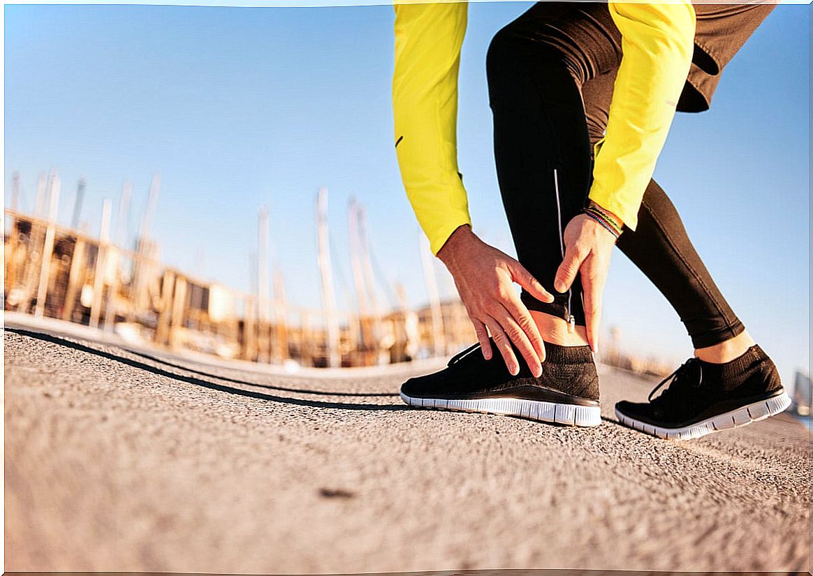
Twin Fibrillar Rupture: Symptoms, Causes And Treatment
Fibrillar rupture is an alteration that affects groupings of fibers that form muscular structures. In the muscles we also find other types of tissue (blood vessels, connective tissue and nerve connections, among others).
It usually occurs more frequently in the lower extremities (especially in the thigh and calf area), although it can also appear in other regions of the body.
Symptoms of a twin fibrillar tear
After the fibrillar rupture has occurred, during the first moments the person may not feel any discomfort, but as time progresses, the first symptoms begin to appear. It should be noted that the severity of the symptoms will depend on the number of fibers that have been injured.

Among the most common symptoms we can highlight:
- Severe pain or discomfort that begins spontaneously that patients may describe as a stab, a needle stick, and even a stone. This affliction is very localizable, that is, the person is able to pinpoint the area of the tear.
- Difficulty carrying out certain activities involving the damaged area. Therefore, there is a reduction in the mobility of that limb.
- Sensation of resistance or tension in the injured area.
- Presence of bruises or bruises in the affected area due to the rupture of blood vessels in the altered muscle. In milder lesions it does not appear frequently (the connective tissue structure prevents it). However, in the most severe clinical cases the patient usually shows a series of bruises.
- Dizziness, headache, and feeling tired or weak due to discomfort.
Causes of fibrillar tear of the twin
Fibrillar rupture is usually due to an inappropriate attitude when practicing sports. For example, it appears most often in the following situations:
- Failure to perform adequate or insufficient preheating.
- Not having done stretching.
- Carrying out extreme efforts without proper planning or when the muscles are overloaded. A very common case is that of sprinting or practicing sports such as squash.
However, there are other risk factors that increase the chances of suffering from this condition. For example, the use of inappropriate footwear for the practice or the bad conditions that the environment presents to carry out physical activity.
On the other hand, foot deformities (and other orthopedic alterations) together with other diseases that deteriorate the patient’s health status (for example, a cold or the flu) also increase the risk of developing this disorder.
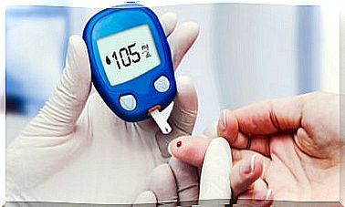
Although in most clinical cases the condition appears in athletes, it can also occur outside the sports field. Certain conditions such as a sedentary lifestyle, an inadequate diet and metabolic diseases (such as diabetes) weaken the general muscles.
Diagnosis and treatment
In order to make the diagnosis, a physical examination of the affected area is performed and, additionally, some internal imaging tests are indicated. The most used are the following:
- Ultrasound scans.
- Computerized axial tomography (CT).
- Magnetic resonance imaging (MRI).
It should be noted that internal imaging tests not only detect the break but also allow its evolution to be verified.

When it comes to treatment, physical activity should be stopped completely first. Next, a compression bandage should be placed and the application of local cold (if ice is used, it should not be applied directly to the skin). In this way, inflammation and severe pain can be reduced.
Additionally, the doctor may recommend some non-steroidal anti-inflammatory drugs and progressive rehabilitation. This process includes performing gentle stretches once the severe discomfort has subsided.
In many cases of muscle strain, physical therapy can be an excellent complement to the treatment prescribed by the doctor. This is because it allows to alleviate discomfort, work delicately on the mobility of the area, to avoid the consequences of complete rest, and facilitate recovery.
On the other hand, the professional might consider authorizing kinesiotaping or neuromuscular bandaging as a complementary measure for relief.